The collection of the Anatomical Museum

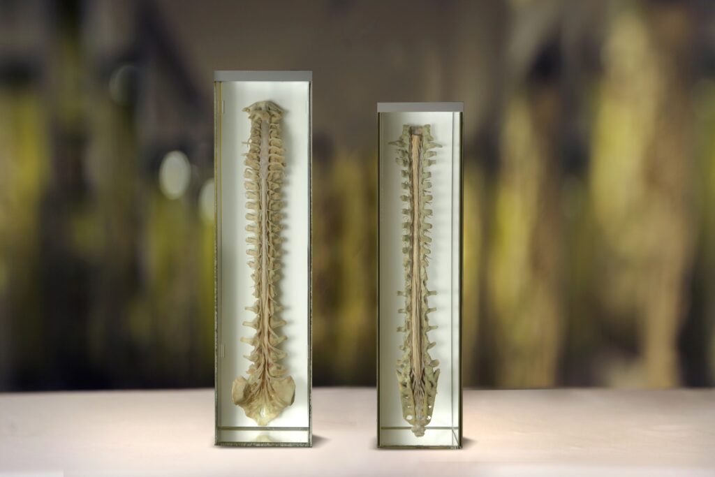
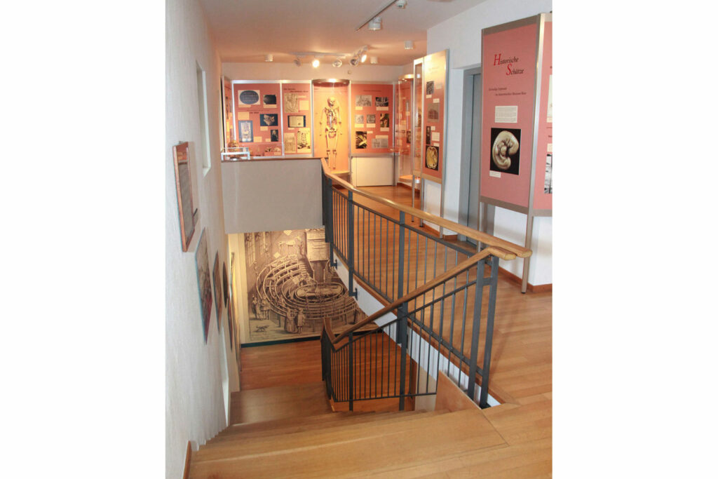
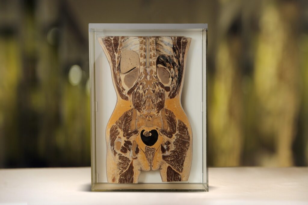
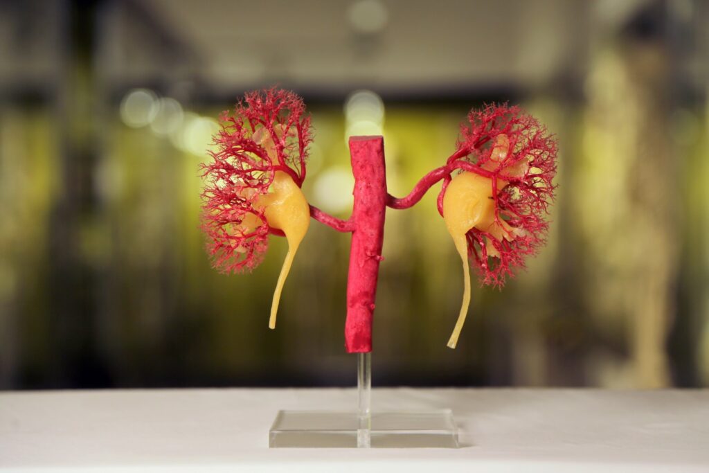
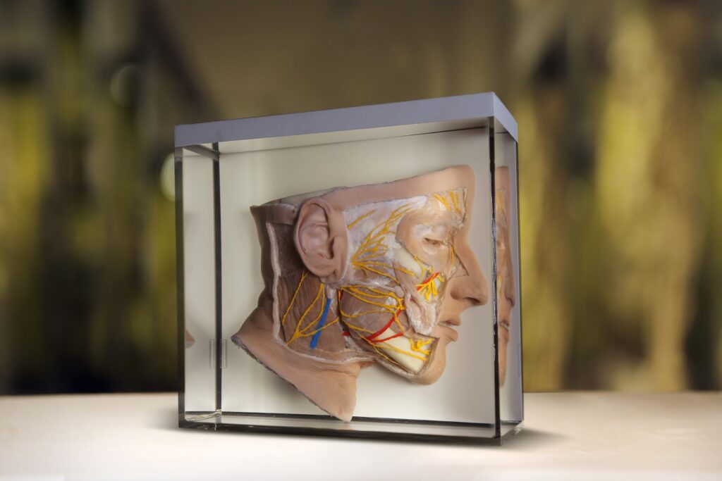
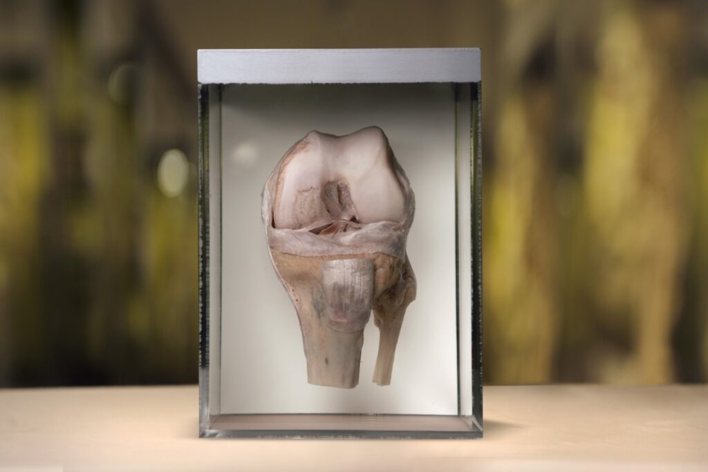
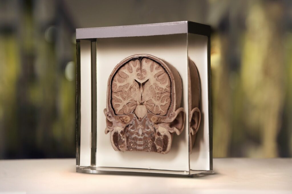
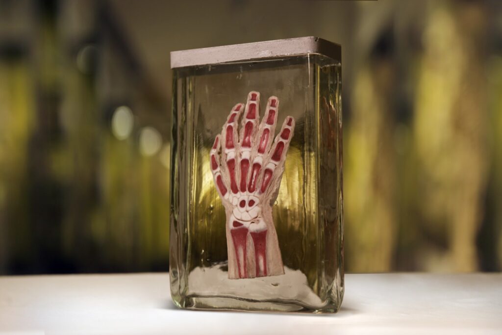
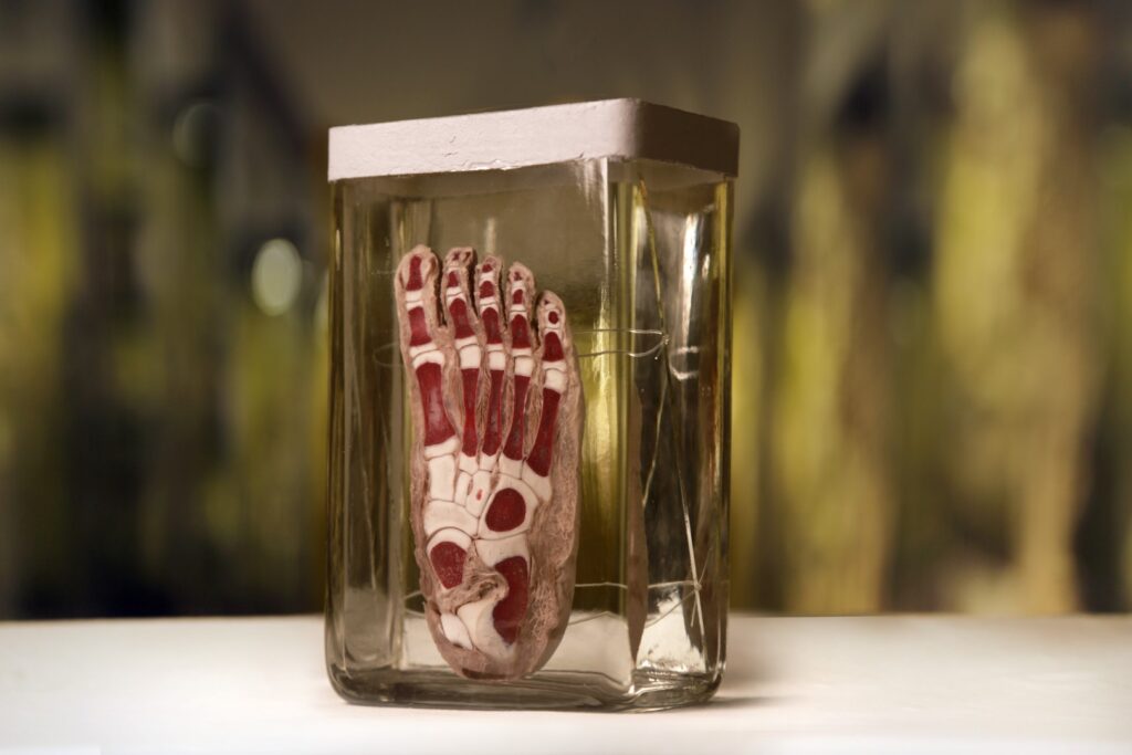
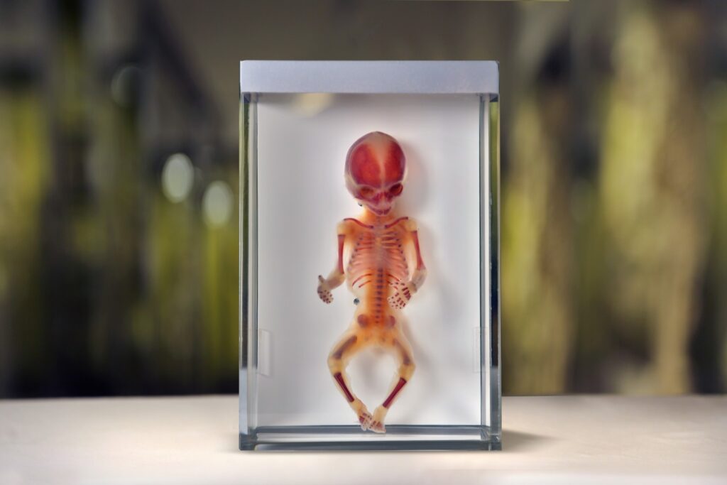
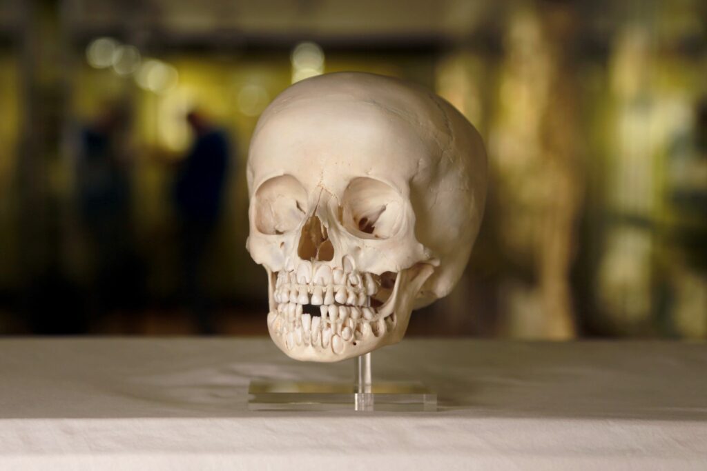
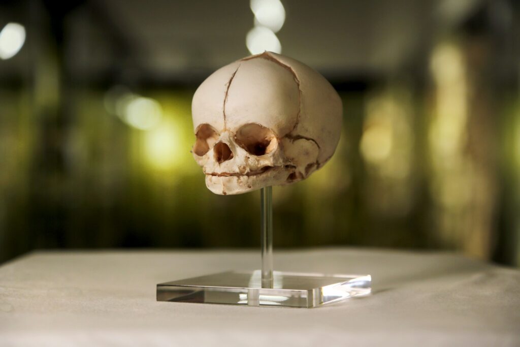
The Anatomical Museum of the University of Basel was founded by Prof. Carl Gustav Jung in 1824. Original preparations of human body parts, organs and tissue are displayed in the museum. They are shown in a systematic and topographic order and describe the structure of the human body. Exhibits of prenatal development are also shown. Special exhibitions explain specific areas of anatomy in a way, which is understandable for all visitors. The museum houses contemporary exhibits as well as numerous historically valuable preparations, which were restored using modern techniques and are now shown in new displays. Of particular interest is the skeleton, which was prepared by Andreas Vesale in 1543 in Basel. It is known as the oldest anatomical preparation of a skeleton in the world. Also of interest is a preparation made in 1573 by Felix Platter, wax models made in 1850 by Carl Gustav Jung and body cross-sections (slices) made in 1900 by Hanson Kelly Corning.


  • left: Medulla spinalis,
    Anterior spinal canal opened,
    adult, Th. Keller/K. Strasser 1935

    right: Medulla spinalis
    Spinal canal opened from behind
    Adult, E. Kupper 1929
  • Stations for listening and understanding.
  • Capturing anatomy in 3D.
  • Anatomy divided into the different organ systems.
  • Historically significant preparations from 5 centuries.
  • Frontal section of the torso

    Section 4, adult section series, 1900
  • Renal pelvis

    Pelves renales (yellow)
    Dendritic-ampullary mixed form
    Renal arteries red
    View from behind
    Corrosion preparation
    Adult
    H. Short 1980
  • Arteries and nerves of the face

    yellow: facial nerve,
    branches of the mandibular nerve
    red: branches of the external carotid artery
    Adult
    A. Wolf 1952
  • Left knee joint

    Representation of the cruciate ligaments,
    collateral ligaments and menisci
    in a flexed position,
    View from the front,
    Adult 1949
  • Frontal section of the head

    Section 5, section series 1 year old child, 1904
  • Hand

    Longitudinal section, bone red
    dyed, approx. 3-year-old child
    1932
  • Foot

    The bone substance is stained red.
    Approximately 2-year-old child.
    1932 Rest. 1980
  • Dies ist der Titel für Bild 15

    Seat height (SSL): 12.0 cm
    Total size: 19.5 cm
    Age: 18. week
    Bone substance stained red
    Rest. 1995
  • Teeth change

    The anlagen of the permanent teeth
    are exposed. Skull of an approx.
    6-year-old child.
    1923 Rest 1985
  • Skull

    Cranium. Newborn.
    Rest. 1995

Rundgang


Anatomisches Museum
der Universität Basel
Pestalozzistrasse 20
CH - 4056 Basel

+41 061 207 35 35
museum-anatomie@unibas.ch

Opening hours:

Monday to Friday 14.00 to 17.00 hours
Sunday 10.00 to 16.00 hours
Sa and general holidays closed
chevron-upchevron-leftchevron-right